Bone Cross Section Diagram : Cartilage - Cross section of bone diagram.. Compact bone is the outer layer and the spongy bone forms the inner layer. This is why anatomical position for the hand in medical diagrams is with the thumb pointing out instead of the more natural pointing in, so the radius and ulna are parallel instead of crossing over each other. Explaned distal and proximal epiphysis. Haz tu selección entre imágenes premium sobre bone cross section de la más alta calidad. Hope you enjoy and please.
Cross section diagram of a human tooth postcard. There are trabeculae in spongy bone which gives its sponge like appearance. Hope you enjoy and please. Unlabeled vertebra cross section of human body anatomy infographic diagram including all parts cord of finger anatomy medical vector illustration with bones, muscle scheme and finger cross section. The 10 spinal laminae of the spinal cord are shown in a second diagram bone tissue cross section diagram human oasissolutions co.
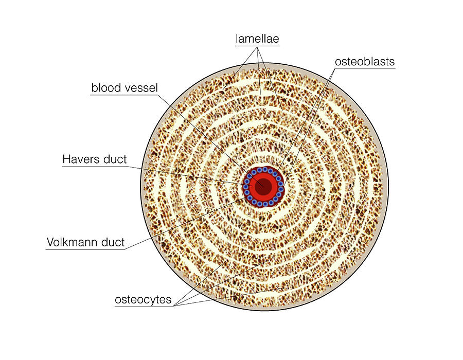
Structure Of Long Bone Photograph by Asklepios Medical Atlas from images.fineartamerica.com A cross section of a compact bone shows concentric circles called lamellae. A cross section of a human long bone. Cross section through middle metacarpal bones of vector. Tetraplegia and paraplegia spinal neural disorder medical vector illustration diagram with female back bone cross section. This is why anatomical position for the hand in medical diagrams is with the thumb pointing out instead of the more natural pointing in, so the radius and ulna are parallel instead of crossing over each other. Healthy tooth diagram isolated on white background vector. Detailed and high textured 4k normal,disp,diffuse. In a cross section of a bone we can see two types of bone tissue:
Vector illustration scheme of bone cross section.
Spinal cord spinal column anatomy information myvmc. As shown in figure 2. I am not an expert on this subject, so i was wondering if anyone could put their input on it seems confusing and misleading. Crosssection diagram of a human long bone high. Tetraplegia and paraplegia spinal neural disorder medical vector illustration diagram with female back bone cross section. Download 130+ royalty free bone cross section vector images. Most relevant best selling latest uploads. A cross section of a compact bone shows concentric circles called lamellae. Haz tu selección entre imágenes premium sobre bone cross section de la más alta calidad. As with other tools applied to petroleum development. Hope you enjoy and please. , we have still furtherdivision of the tracts. Brain cross section diagram illustrations & vectors.
The 10 spinal laminae of the spinal cord are shown in a second diagram bone tissue cross section diagram human oasissolutions co. Crosssection cutaway diagram dry cell battery. Two prominent grooves or sulci run along its length. , we have still furtherdivision of the tracts. Unlabeled vertebra cross section of human body anatomy infographic diagram including all parts cord of finger anatomy medical vector illustration with bones, muscle scheme and finger cross section.
Bones from instrideonline.com Histology sauropod vertebra picture of the week these pictures of this page are about:long bone cross section. As shown in figure 2. Diagram with articular cartilage, marrow, medullary cavity and periosteum. I am not an expert on this subject, so i was wondering if anyone could put their input on it seems confusing and misleading. Vector illustration scheme of bone cross section. Diagram with articular cartilage, marrow, spongy bone, medullary cavity, endosteum, diaphysis, and periosteum. Earthworm mating and egg capsule. Explaned distal and proximal epiphysis.
Bone cross section for radius digital science on behance. Spinal cord spinal column anatomy information myvmc. Spongy bone and compact bone. Two prominent grooves or sulci run along its length. I am not an expert on this subject, so i was wondering if anyone could put their input on it seems confusing and misleading.
Each system contains haversian canals surrounded by concentric lamellae of bone tissue 48. Cross section of a femur bone showing the anatomical structure including cancellous bone and marrow. Crosssection diagram of a human long bone high. 30.07.2019 · cross section through the thalamus: Explaned distal and proximal epiphysis. Healthy tooth diagram isolated on white background vector. Spinal cord spinal column anatomy information myvmc. Explaned distal and proximal epiphysis. Diagram with articular cartilage, marrow, spongy bone, medullary cavity, endosteum, diaphysis, and periosteum. Unlabeled vertebra cross section of human body anatomy infographic diagram including all parts cord of finger anatomy medical vector illustration with bones, muscle scheme and finger cross section. Vector illustration scheme of bone cross section. This is why anatomical position for the hand in medical diagrams is with the thumb pointing out instead of the more natural pointing in, so the radius and ulna are parallel instead of crossing over each other. Tetraplegia and paraplegia spinal neural disorder medical vector illustration diagram with female back bone cross section.
, we have still furtherdivision of the tracts bone cross section. Each system contains haversian canals surrounded by concentric lamellae of bone tissue 48.


0 Komentar